Fig. 2.

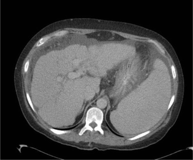
Portal venous CT demonstrates an irregular nodular liver, with the capsule outlined by ascites. Note splenomegaly due to portal hypertension

Portal venous CT demonstrates an irregular nodular liver, with the capsule outlined by ascites. Note splenomegaly due to portal hypertension