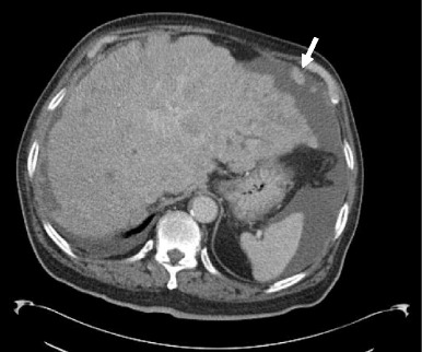
Fig. 6.

Portal venous phase CT demonstrates an enlarged liver with a markedly irregular contour due to metastatic melanoma. The patient has ascites and peritoneal deposits (arrow), and clinically was heavily jaundiced, with gross hepatic dysfunction. Note normal-sized spleen
