
Fig. 2.
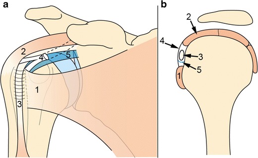
Schematic representation of the rotator interval (RI): front view (a), sagittal view (b). The RI is bordered by the SSC tendon inferiorly (1) and the SSP tendon superiorly (2) and contains the intra-articular portion of LHBT (3). CHL (4) and SGHL (5) surround the LHBT in the RI, acting as a pulley system for LHBT stabilization