
Fig. 3.
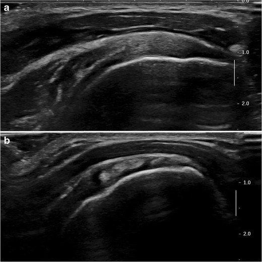
B-mode ultrasound images of the SSC. a Transverse view (long axis) : the normal SSC tendon is hyperechoic compared to the adjacent muscle and has a fibrillary structure. b Sagittal view (short axis): multi-pennate structure of the SSC tendon with alternation of hypo and hyperechoic zones