
Fig. 4.
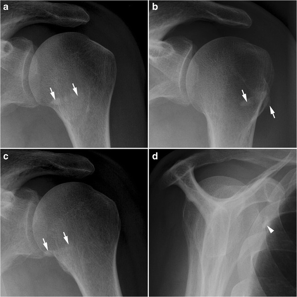
Calcific tendinitis of the SSC. Calcificationsof the SSC tendon (white arrows) overlap the lesser tuberosity in the front view in neutral rotation (a), are lateralized in external rotation (b), and medialized in internal rotation (c). On the Y view, calcifications of SSC tendon are seen under the coracoid process (white arrowhead) (d)