
Fig. 5.
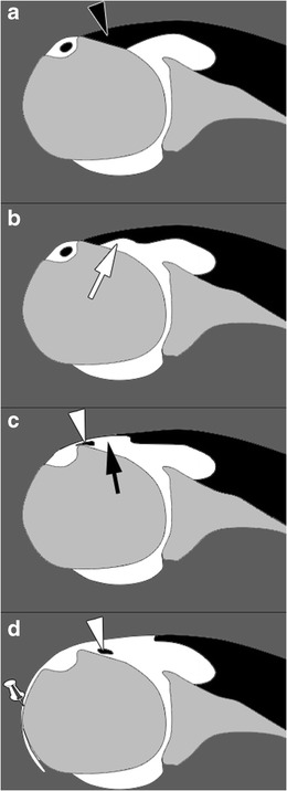
Schematic representation of SSC tendon tears on magnetic resonance or computed tomography arthography. Normal SSC tendon (black arrowhead) (a). Incomplete partial thickness tear (white arrow) (b). Complete partial thickness tear (black arrow) associated with LHBT (white arrowhead) subluxation (c). Complete full thickness tear associated with LHBT (white arrowhead) dislocation and opacification of the SASD bursa (pin) (d)