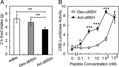
Fig. 4.
Increased activity of Act-αMSH in transfected cells and in vivo.(A) Rats with cannulae preimplanted into the third ventricle were injected with saline or 3 nmol of Des-αMSH or Act-αMSH. Shown is 2 h of food intake. Data are means ± SE of six animals. Significant differences were determined by Student's t test. **, P < 0.005. (B) 293T cells were transiently transfected with MC4R and pCRE-LUC plasmids. Cells were incubated with different concentrations of Des-αMSH or Act-αMSH for 5 h, and luciferase activity was measured. Data are means ± SE of results obtained in triplicate. Significant differences were determined by ANOVA and Student's t test for post hoc analysis. *, P < 0.05; ***, P < 0.0005.

