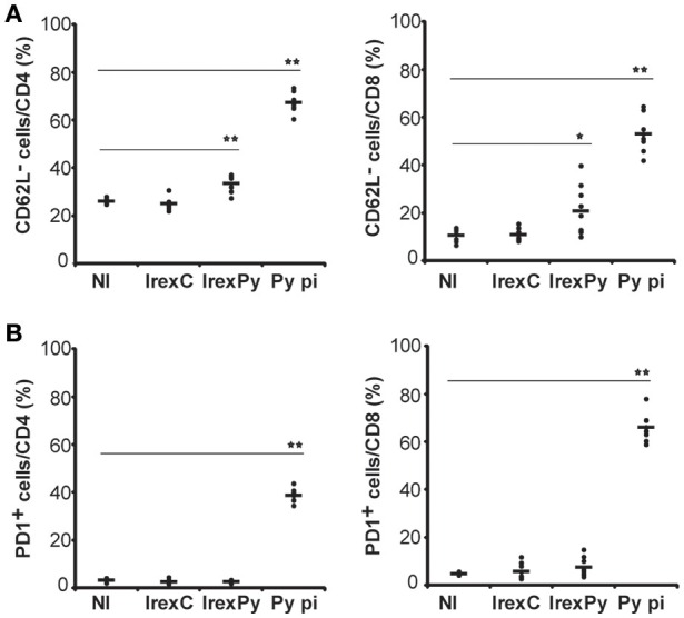
Figure 2.

Analysis of spleen T-cells after immunization with exosomes. BALB/c mice were immunized with rexC or rexPy plus CpG-ODN-1826. Two weeks after the second immunization spleen cells were obtained. Non-immunized mice (NI) were untreated. A group of mice was infected with P. yoelii 17X and spleen cells were obtained 25 days post infection (Py p.i). Spleen cells were labeled with the panel of antibodies described in materials and methods and analyzed in LSRFortessa. (A) Quantification of the percentage of CD4+ or CD8+ CD62L− cells. (B) Quantification of the percentage of CD4+ or CD8+PD1+ cells. (A,B) Bold line corresponds to the mean of 6 to 9 mice per group from three different experiments. Percentages are evaluated with analysis of variance and vs.non-immunized group (Dunnet post hoc test). (*P < 0.05, **P < 0.01).
