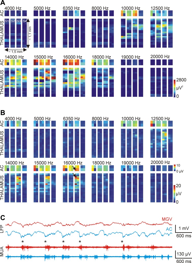
Fig. 10.
Tonotopic mapping in the ventral division of the auditory thalamus of the anesthetized rat. Tonotopic maps were constructed from the integral (A) and the peak value (B) of the first 100-ms-long section of the mean evoked MUA response to acoustic stimulation (n = 20 trials). Pure tones with 12 different frequencies (4–20 kHz) were used for tonotopy. The construction of tonotopic maps was done in the following way: 4 recording sites had fixed cortical positions on each shaft, whereas the remaining 4 electrodes were located in the thalamus. After a few minutes of acoustic stimulation and continuous recording of the neural activity, thalamic sites were repositioned. Using this procedure, we could map the sound-evoked brain activity on the section of the recording probe that was located in the thalamus. C: local field potential (LFP) and MUA traces corresponding to a cortical and a thalamic site (indicated with black arrows in B) with a match in the tonotopy at 16 kHz. The asterisks indicate up-states with synchronous onset. AC, auditory cortex; MGV, ventral medial geniculate.

