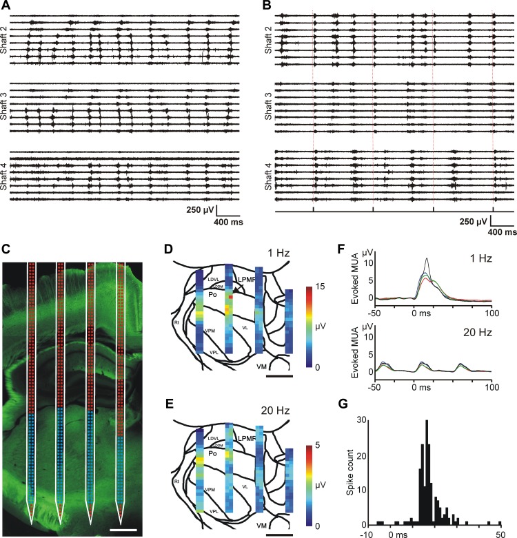
Fig. 11.
Optogenetic experiments with the EDC probe in mice. A: 4-s-long spontaneous MUA traces recorded under ketamine-xylazine anesthesia in the mouse thalamus. Traces recorded on 3 shafts of the EDC probe are shown. Note the different propagation patterns of thalamic up-states across recording sites. The electrodes were configured in a way similar to that shown in Fig. 8C (expanded configuration). These propagating patterns bear a close resemblance to thalamic up-state propagation we observed in rats (see Fig. 8, C and D). B: 4-s-long MUA traces recorded in the thalamus of the anesthetized mouse during optical stimulation. In this case octrode configuration was used for MUA recordings. Optical stimulation of the somatosensory cortex (0.8 mW, 1 Hz) elicited MUA responses in the thalamus. The delivery times of the laser stimuli are shown on the trigger channel (bottom) and are also indicated with dashed red lines. Strong MUA response is present on shaft 2 after each stimulus. C: fluorescent light microscopic image of the enhanced yellow fluorescent protein (EYFP) signal in the thalamus at the site of the probe penetration with the schematic of the probe overlaid on the photomicrograph. The location of the probe was estimated on the basis of the position of the probe tracks and the implantation depth. The activity recorded on electrodes with blue color was used to construct color maps shown in D and E. Scale bar, 500 μm. D and E: color maps showing the peak amplitude of the mean MUA response at every thalamic recording site in response to optical stimulation of the somatosensory cortex with 1-Hz (D) and 20-Hz (E) frequencies. Sites with no detectable MUA were excluded from the analysis. Scale bar, 500 μm. F: the averaged (n = 30 trials) and filtered (100 Hz, low-pass filter) MUA response to 1-Hz (top) and 20-Hz (bottom) blue light stimulation recorded on 4 adjacent electrodes. The position of recording sites is indicated with a black arrow in D. Optical stimuli were delivered at time 0. G: peristimulus time histogram (PSTH; optical stimuli vs. spike times) of a light-responsive thalamic single unit (bin width: 1 ms). Optical stimuli (n = 390) were delivered at time 0.

