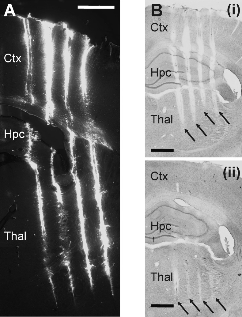
Fig. 4.
Postmortem histological reconstruction of the probe tracks to determine the location of the EDC probe in the rat brain. A: fluorescent micrograph of a coronal rat brain section containing the tracks of the 4 shafts of the EDC probe. The back side of the shafts was painted with DiI before the acute experiment. The tip of the shafts can be clearly located. Scale bar, 1 mm. B: light microscopic image of two 60-μm-thick Nissl-stained coronal rat brain sections showing the 4 tracks of the probe. The 2 sections displayed are not adjacent; there was an additional section located between them (not shown). Black arrows point to the tracks made by different shafts. The reason for the visibility of the tracks on multiple sections is presumably due to the oblique cutting of the fixed brain before staining. Scale bar, 1 mm. Based on the tracks visible on Nissl-stained sections and the known penetration depth, the approximate position of recording sites can be determined, together with the main structures from which the neuronal activity was recorded. Ctx, neocortex; Hpc, hippocampus; Thal, thalamus.

