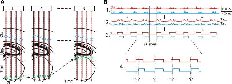
Fig. 6.
Recording the thalamocortical slow-wave activity (SWA) with the EDC probe in multiple brain areas and the steps used to process the recorded data. A: mapping protocol used to record simultaneous activity in the thalamus and the neocortex. At least 2 recording sites on each shaft were selected in the cortex where unit activity with high SNR was detected (blue circles). The remaining recording sites were positioned near the tip of the probe, which was located in the thalamus (green circles; 1). After a few minutes of continuous recording, these thalamic sites were repositioned to new sites in the thalamus and a new recording file was started (2). This procedure was continued until the whole thalamus was mapped (N). B: multiple-unit activity (MUA)-based detection of up- and down-state onsets. After the wideband data were filtered (bandpass filter, 500-5,000 Hz; 1), the envelope of the MUA signals was obtained with a low-pass filter (30 Hz; 2). A threshold level was calculated on each channel based on the average and SD of MUA during down-states (ThCtx and ThThal). The calculated threshold was used to acquire the up- and down-state onsets (3). Finally, the time difference between cortical and thalamic up-state onsets was calculated by subtracting the time point of the cortical up-state onset from the time point of the thalamic up-state onset (4). Thalamic traces are red and cortical traces are blue.

