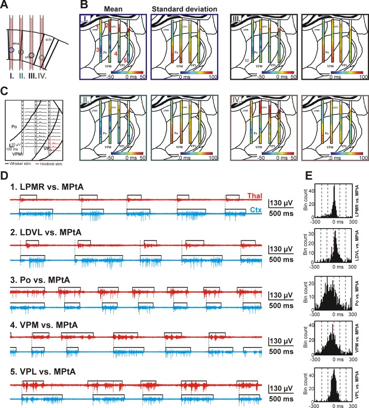
Fig. 7.
Spatiotemporal dynamics of the thalamocortical SWA observed in a representative rat. A: the 4 positions (1 per shaft) from where cortical recordings were obtained and used to derive the color maps shown in B (I-IV). Recording sites were located in layer 5 of association and somatosensory cortices. The relationship between the cortical sites and the color maps in B is color coded. B: color maps constructed from the mean and SD of the time difference of up-state onsets calculated between the cortical sites shown in A and between various thalamic sites. The color maps are representative examples from a single rat. On the color map of the mean up-state onset time difference, blue colors indicate that up-states detected on thalamic sites usually started before cortical up-states, whereas red colors represent the lead of cortical up-states. White color at the top of the color map indicates areas where no significant MUA could be detected. C: in addition to histological verification, the thalamic position of recording sites was also verified using somatosensory stimulation. In this representative example, repetitive (∼1 Hz) whisker (black) and hindlimb stimulation (red) was applied to determine the approximate position of the borders between VPL, VPM, and Po thalamic nuclei. Evoked MUA responses on 3 shafts are shown. D: representative MUA traces showing the up-state dynamics between different thalamic and cortical sites. The position of thalamic sites is indicated in B next to the mean color map corresponding to cortical site I (top left; Arabic numerals with red color). Black rectangles show the detected up-states. The onset of cortical up-states is marked with dashed black lines. E: histograms of up-state onset time differences calculated between recording sites presented in D. LDVL, laterodorsal ventrolateral nucleus; LPMR, mediorostral part of the lateral posterior nucleus; LPtA, lateral parietal association cortex; MPtA, medial parietal association cortex; Po, posterior nucleus; S1Tr, trunk region of the primary somatosensory cortex; VPL, ventral posterolateral nucleus; VPM, ventral posteromedial nucleus.

