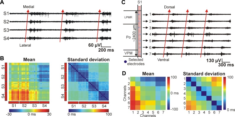
Fig. 8.
Preferred cortical and thalamic propagation patterns of thalamocortical SWA under ketamine-xylazine anesthesia. A: 2-s-long MUA traces of 4 cortical recording sites (S1–S4) located on different shafts showing 3 consecutive up-states recorded in layer 5. The recording sites were selected near the sites indicated in panel A of Fig. 7. The up-states propagated most frequently from the most lateral shaft ('S4', primary somatosensory cortex) in the direction of the most medial shaft ('S1', medial parietal association cortex) with a latency of 10–30 ms (red arrows). B: Mean (left) and standard deviation (right) of the up-state onset time difference between up-states detected on cortical recording sites located in layer 5. The color maps were constructed from the data set of the same animal shown in A and in Fig. 7. However, in this case all recordings sites were selected in the cortex. On the color map of the mean up-state onset time difference, red color represents the lead of up-states detected in positions located on the y-axis relative to the positions on the x-axis. C: 3-s-long MUA traces showing the ventral-to-dorsal propagation of up-states detected in the thalamus (red arrows) on 1 shaft of the EDC probe. The latency between the up-state onsets detected on the most ventral and on the most dorsal electrode was greater than 50 ms. The 7 thalamic recording sites were selected in a way to be able to record from a larger volume of thalamus in the dorsoventral plane, covering several nuclei (left). The eighth site was located outside the thalamus. D: mean (left) and SD (right) of the up-state onset time difference between up-states detected on thalamic recording sites. The recording sites were selected on that shaft that was located most medially. On the color map of the mean up-state onset time difference, red color represents the lead of up-states detected in positions located on the y-axis relative to the positions on the x-axis.

