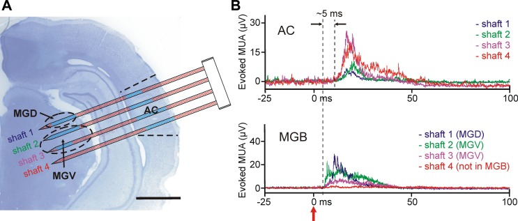
Fig. 9.
Electrical activity recorded with the EDC probe from the auditory thalamocortical system of the anesthetized rat. A: the schematic of the probe overlaid on the top of a Nissl-stained coronal rat brain section containing the probe tracks. The shafts of the probe were located in the auditory cortex (AC) and in the ventral (ventral medial geniculate, MGV) and dorsal (dorsal medial geniculate, MGD) divisions of the medial geniculate body (MGB). To penetrate through both structures, the probe was inserted at an angle of 70° relative to the dorsoventral axis. After the implantation of the probe, click stimuli were delivered to the contralateral ear of the animal and the electrical activity was examined at every electrode position that was located in the brain, to find areas responding to sound. Blue dots show recording sites with significant MUA response to click stimuli. Scale bar, 2 mm. B: representative mean evoked MUA responses (averaged from n = 20 trials) in the AC (top) and in the MGB (bottom) to acoustic click stimulation. A single example from each shaft is presented. There was no response to sound on that part of the fourth shaft, which was located in the thalamus. The click stimulus was delivered at time 0 (red arrow).

