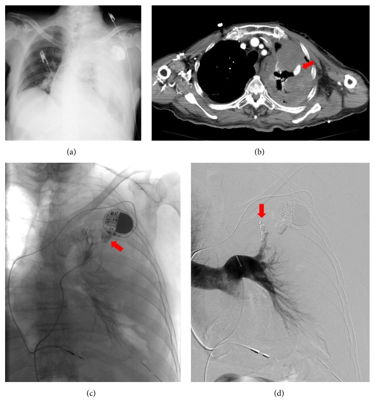
Figure 1.
A 90-year-old man received a permanent implanted pacemaker. After the procedure, the patient's blood pressure steadily decreased. Left hemothorax and laceration of the pulmonary arterial branch of the left upper lobe were confirmed. (a) Chest radiograph showed a left hemothorax. (b) Contrast-enhanced computed tomography (CT) showed a confined cavity (pseudoaneurysm) of a branch of the upper lobe pulmonary artery (arrow). (c) Angiography demonstrated bleeding from the perforated pulmonary arterial branch of the left upper lobe (arrow). (d) Transcatheter arterial embolization (TAE) was performed using microcoils (arrow). Digital subtraction angiographic image taken after coil embolization discloses coils (arrows) in the upper lobe pulmonary artery and no further opacification of the pseudoaneurysm.

