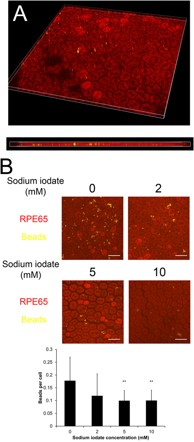
Figure 5. The effect of sodium iodate on RPE phagocytotic activity.

Photoreceptor outer segment phagocytosis was performed on the rat RPE explant culture. RPE cells were visualized by the immunofluorescence signal of RPE65 (Red), whereas FITC-labeled latex beads with rat POS (yellow) were opsonized. (A) The confocal microscopy image of the phagocytosed, POS opsonized and FITC-labeled latex beads in the RPE65-stained RPE cells. (B) After 5-day sodium iodate treatment, the phagocytotic activity of RPE cells was dose-dependently attenuated from 2–10 mM sodium iodate. Scale bars: 50 μm. ‘**’p < 0.01
