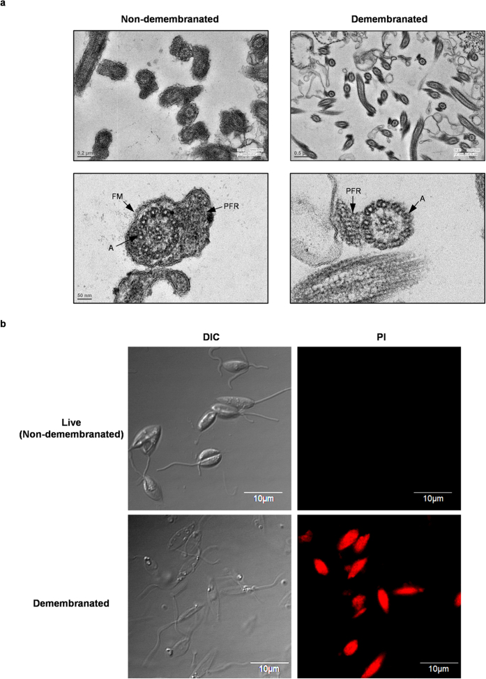
Figure 1. Permeabilization of L. donovani by 0.1% Triton demembranation.
(a) Transmission electron micrographs (TEMs) of cross-sections of flagella of live and demembranated L. donovani. In live (non-demembranated) Leishmania intact outer flagellar membrane (FM) is visible. In demembranated Leishmania outer flagellar membrane (FM) is absent due to extraction with 0.1% Triton. (A) axoneme and (PFR) paraflagellar rod. (b) Confocal microscopy images of live (non-demembranated) and demembranated L. donovani treated with 15 μM propidium iodide (PI). Images were captured at 100X using an oil-immersion objective.

