
Figure 3.
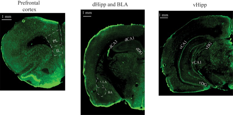
Representative images in the vmPFC, dHipp, BLA, and vHipp obtained after performing c-Fos immunocytochemistry and scanning brain slides using the Li-cor Odyssey scanner. (vmPFC) ventromedial prefrontal cortex, (IL) infralimbic cortex, (PL) prelimbic cortex, (dHipp) dorsal hippocampus, (BLA) basolateral amygdala, (vHipp) ventral hippocampus.