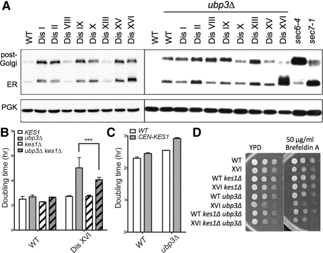
Figure 2.

Deletion of UBP3 enhances the vesicle transport defect of disome XVI cells. (A) Cells were grown to mid-log phase in YPD at room temperature. Ccw14 mobility was analyzed by Western blot analysis. The ER and post-Golgi forms of the protein are indicated. Known secretory mutants sec6-4 and sec7-1 are shown for comparison. Pgk1 was used as a loading control. The lysates for the blot at the left (disomes wild type [WT] for UBP3) are from Dodgson et al. (2016). (B,C) Doubling times of the indicated strains were determined as described in Figure 1. (***) P < 0.0001, Student's t-test. (D) Cultures of the indicated strains were grown overnight in YPD, and 10-fold serial dilutions were plated on YPD plates with or without the indicated concentration of Brefeldin A at 30°C.
