Figure 1.

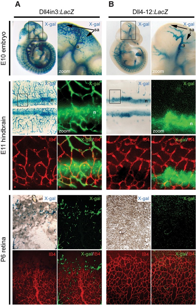
The Dll4in3 enhancer directs gene expression to endothelial cells during sprouting angiogenesis. (A) Representative images from Dll4in3:LacZ transgenic mice demonstrate enhancer activity in endothelial cells undergoing sprouting angiogenesis in the E10 embryo, E11 hindbrain, and postnatal day 6 (P6) retina. (B) Representative images from Dll4-12:LacZ transgenic mice demonstrate enhancer activity in arterial and neural tissues but no activity in endothelial cells during sprouting angiogenesis in E10 embryos, E11 hindbrains, or postnatal retinas. Enhancer activity was detected as X-gal activity (blue staining or green pseudocolor), and endothelial cells were detected by isolectin B4 (IB4) whole-mount immunostaining (red). (a) Artery; (sa) region of sprouting angiogenesis; (n) neuronal staining. See also Supplemental Figure 1.
