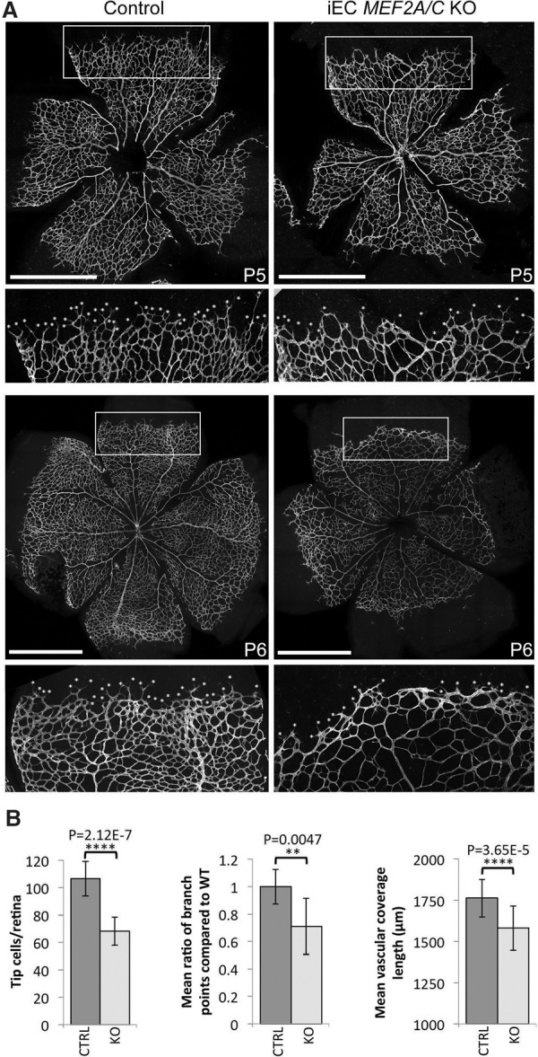
Figure 5.

Induced endothelial deletion of Mef2A and Mef2C results in reduced retinal angiogenesis. (A) Representative P5 and P6 retinas taken from control (Cre-negative) and iEC Mef2A/C knockout (KO) pups and stained for IB4. Tip cells were detected by filopodia and are indicated by asterisks. The white box indicates region taken for zoom image. Bars, 1 mm. (B) Graphs demonstrating the mean number of tip cells, the ratio of the mean number of branch points, and the mean outgrowth length as measured by actual distance covered by the growing vessel from the center of the retina. Statistical analysis was performed on seven wild-type and nine knockout pups pooled from two different litters.
