Figure 7.
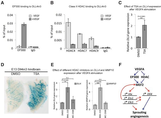
VEGFA signaling leads to activation of MEF2 transcriptional activity. (A) Increased EP300 binding at the DLL4in3 enhancer after VEGFA stimulation in HUVECs analyzed by ChIP. Graph is representative of four biological replicates. (B) Decreased class II HDAC binding at the DLL4in3 enhancer after VEGFA stimulation in HUVECs analyzed by ChIP. The graph is representative of two biological replicates. (C) Relative DLL4 gene expression after VEGFA stimulation with and without trichostatin A (TSA), analyzed by qRT–PCR. Statistical analysis on four biological replicates. Error bars indicate standard deviation. (D) Representative Dll4in3:LacZ E13 hindbrains removed 24 h after in utero intracerebral injection of 10 μM HDAC inhibitor TSA or DMSO control. X-gal staining after DMSO control injection is weak, whereas more robust transgene expression was detected in hindbrains injected with TSA. (E) Relative gene expression levels of DLL4 and MMP10 after VEGFA stimulation with and without treatment with small molecule class II HDAC inhibitors BML-210 and MC-1568, analyzed by qRT–PCR. Statistical analysis was performed on three biological replicates. Error bars indicate standard deviation. (F) Proposed model of VEGFA-mediated activation of sprouting angiogenesis via the MEF2 transcription factor family. See also Supplemental Figure 9.

