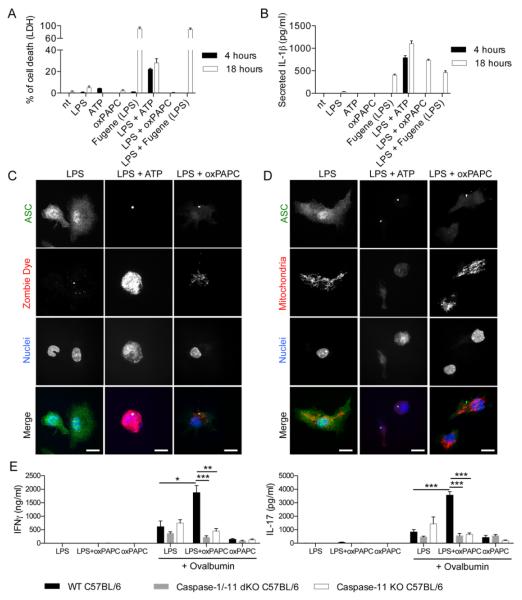
Fig. 4. oxPAPC prevents DC death and potentiates adaptive immune responses.
(A and B) DCs were treated with LPS alone, ATP alone, oxPAPC alone or FuGENE complexed LPS [Fugene (LPS)], or were primed for three hours with LPS and then treated with the indicated stimuli. Cell death was measured by LDH release (A) or IL-1β secretion was measured by ELISA (B). Means and standard deviations of four replicates are shown. (C and D) DCs were pretreated with LPS for 3 hours and then activated with ATP or oxPAPC. 18 hours later, cells were stained for ASC (green), nuclei (blue) Zombie Dye (red) (C) or active mitochondria (red). Scale bar: 10 μm. (D). Panels are representative of three independent experiments. (E) CD4+ T-cells were isolated from the draining lymph nodes 40 days after immunization with OVA + LPS in IFA (LPS), OVA + LPS + oxPAPC in IFA (LPS+oxPAPC) or OVA + oxPAPC in IFA (oxPAPC) of WT, caspase-1/-11 dKO or caspase-11 KO mice. CD4+ T-cells were restimulated or not with OVA in the presence of DCs. IFNγ (left panel) and IL-17 (right panel) secretion was measured 5 days later by ELISA. Bar graphs represent means and standard errors of two experiments with five animals per group. *P < 0.05; **P < 0.01; ***P < 0.005.

