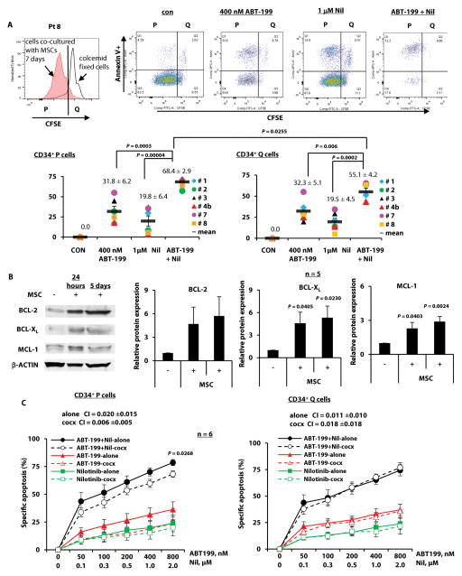
Fig. 6. Targeting of BCL-2 and BCR-ABL in proliferating and quiescent CD34+ cells from TKI-resistant BC CML patients.
(A) CFSE-stained cells were treated with ABT-199, nilotinib, or both. Apoptosis in proliferating (P) and quiescent (Q) CD34+ cells was assessed after 48 hours. Upper panel shows flow cytometric profiles of cells from patient 8 (Pt 8) before and after treatment. Lower panel shows the results of six treated patient samples, where each dot represents the results for one patient sample. (B) Expression of BCL-2, BCL-XL, and MCL-1 in cells from CML patient samples co-cultured with MSCs (cocx) for 24 hours or 5 days was examined using immunoblot. (C) CFSE-labeled cells (Table 1, n = 6) were treated with ABT-199, nilotinib, or both with or without MSC co-culture. Apoptosis was assessed in proliferating and quiescent CD34+ cells after 48 hours. con, control; Nil, nilotinib.

