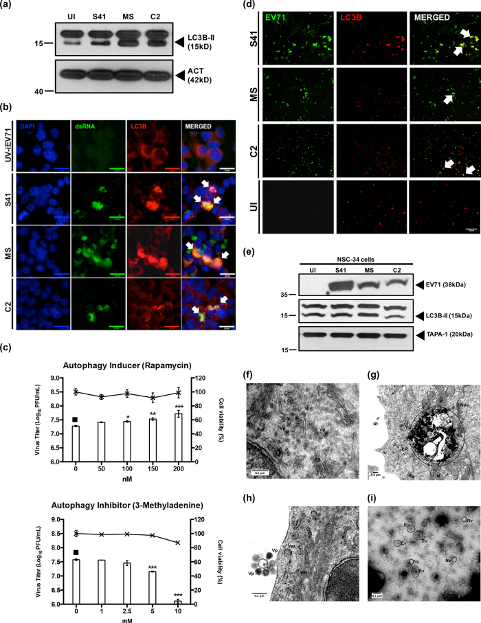
Figure 6. Autophagy in EV71-infected NSC-34 cells and detection of EV71-containing autophagic vacuoles in the culture supernatant.
NSC-34 cells were infected with S41, C2 and MS strains at MOI 10 and at 48 h.p.i. (a) Western blot analysis was performed on the cell lysates using anti-LC3B primary antibodies. β-actin was used as the loading control. Gels images were cropped. (b) Immunostaining using antibodies specific to LC3B protein (red) and dsRNA (green). White arrows indicate co-localization of both signals. Images were taken at 60× magnification. Representative views are shown. Scale bar denotes 20 μm. UV-iEV71 infection served as negative control. (c) NSC-34 cells were treated with autophagy inducer (rapamycin) prior to S41-infection, or were treated post-infection with autophagy inhibitor (3MA). The culture supernatants were harvested at 48 h.p.i. for virus titer determination. Cytotoxicity of rapamycin and 3MA in NSC-34 cells was determined using alamarBlue™ cytotoxicity assay. Data are expressed as the mean ± SD of technical triplicates. Statistical analysis was performed using one-way ANOVA with Dunnett’s post-test (*p < 0.05, **p < 0.005, ***p < 0.0005) against untreated cells (black square). (d) Immunostaining of exosome preparations using anti-VP1/VP0 (green) and anti-LC3B (red) antibodies. Arrows indicate co-localization of both signals. Uninfected cells (UI) served as negative control. Images were taken at 60× magnification. Scale bar denotes 10 μm. Representative views are shown. (e) Western blot analysis of the exosome preparations using anti-LC3B, anti-VP1/VP0 and anti-TAPA-1 primary antibodies. Gels images were cropped. (f–h) Transmission electron microscopy images of S41-infected NSC-34 cells (MOI 20) at 24 and 72 h.p.i. (positive staining). (i) Transmission electron microscopy images of exosome preparation from S41-infected NSC-34 culture supernatant (negative staining). Legend: Nuc, nucleus; M, mitochondria; V, vacuole; ER, endoplasmic reticulum; RC, replication complex; Vp, viral particles; Ev, enveloped virus; Nv, naked virus. Representative images are shown. These experiments were performed twice independently. One representative set is shown.

