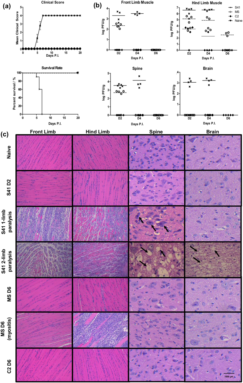
Figure 7. Survival rates, clinical scores, virus titers and histopathology in EV71-infected AG129 mice.
Two-week old AG129 mice were infected with S41, MS or C2 strain at 107 PFU/mouse via the intraperitoneal (i.p.) route. Naive mice received PBS instead. (a) Clinical scores and survival rate (n = 8–10 mice). The mice were monitored over a period of 20 days. Clinical scores were defined as follows: 0, healthy; 1, ruffled hair and hunchback appearance; 2, limb weakness; 3, paralysis of one limb; 4, paralysis of two limbs at which point the animals were euthanized. Data were expressed as mean ± SEM. A p value < 0.0001 was obtained for the log-rank test between S41-infected and naïve mice or C2- or MS-infected mice. Wilcoxon’s test showed that survival curves between S41-infected and naïve or C2- or MS-infected mice were significantly different (p value < 0.0005). (b) Virus titers in front limbs, hind limbs, spinal cord and brain from infected mice (n = 4–5) were determined at day 2, 4 and 6 p.i. by plaque assay. Individual values are shown. Means are represented by solid or dotted lines (refer to legend). (c) Histology analysis. Mice (n = 3) were euthanized at the indicated time points post-infection (MS and C2-infected mice) or upon observation of one or two limbs paralysis (for S41-infected mice). Paraffin sections of front limb, hind limb, spine and brain were stained with H&E and observed under light microscope. Black arrows indicate neuropil vacuolation and neuronal degeneration in the anterior horn region of the spinal cord and brainstem reticular formation of the brain from S41-infected mice. Myositis was observed in one MS-infected mouse at day 6 p.i. All observations were made at 20× magnification. Scale bar denotes 1,000 μm. Representative views are shown. All these experiments were performed two or three times independently. One representative set is shown.

