Figure 1.
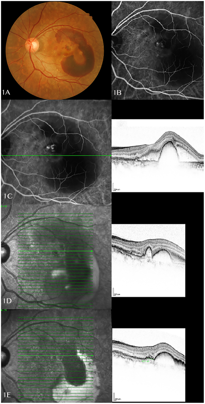
(A) A 57 years old female presented with left eye massive submacular haemorrhage and large pigment epithelial detachments. (B) ICG examination on the same day showed a cluster of polyps at the edge of notched PED, with a kidney-shaped cyanine dye blocked by the submacular haemorrhage. (C) OCT B-scan image showed a central dome-shaped PED with presence of subretinal fluid. Only one OCT image was documented at baseline which shows different sections compared to follow up scans. (D) Repeated OCT examination showed largely subsided subretinal fluid and reduced size of PED at 1 week after the triple therapy treatment. B-scan image showed a reduced size of dome-shaped PED and a continuous small PED corresponding to the polypoidal lesions on ICG. (E) Infrared imaging and OCT B-scan showed further improvement of disease at one month post treatment, especially reduction in the anterior protrusions of a highly reflective RPE line. (ICG: indocyanine green; OCT: optical coherence tomography; PED: pigment epithelial detachment; RPE: retinal pigment epithelium).

