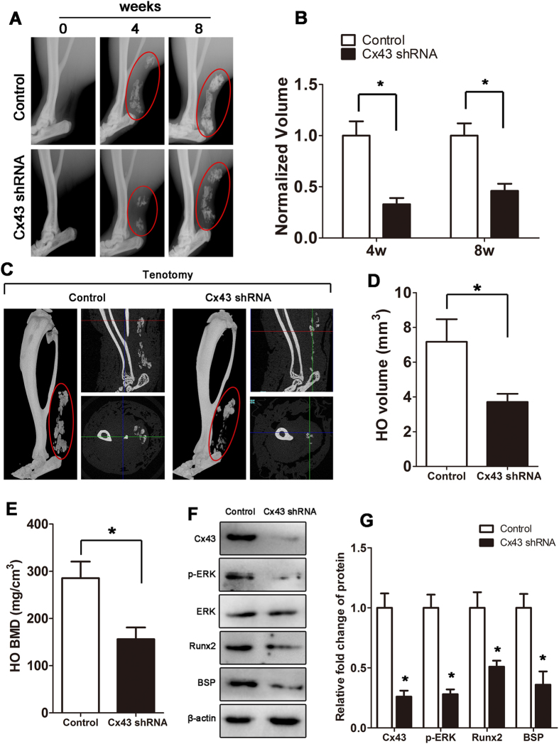
Figure 3. Inhibition of Cx43 limits HO formation in vivo.
Tenotomy mice were injected with Cx43 shRNA or scrambled RNA weekly at the lesion site. (A) HO was visualized by a weekly radiograph. (B) The HO volumes were quantified. (C) At week 8, a three-dimensional reconstruction (Left), cross-section (Right upper) and sagittal section (Right below) of microCT scans of Cx43 shRNA- and control RNA-treated tenotomy mice were obtained. The red, green and blue lines represent the cross, sagittal and coronal plane in other sections, respectively. (D) The HO volumes were quantified. (E) The bone mineral density (BMD) of HO was qualified. (F) Expression of Cx43, p-ERK, ERK, Runx2, BSP and β-actin was examined by Western blot. (G) A densitometric analysis of Cx43, p-ERK, Runx2 and BSP protein expression was performed. (t-test, n = 3, *p < 0.05).

