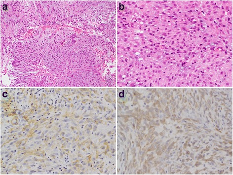
Fig. 2.

Hematoxylin and eosin staining (a, b) and immunohistochemical staining (c, d). a The proliferation of plump spindle cells with a storiform architecture with scattered lymphocytes. Epithelioid components with an appearance suggestive of odontogenic epithelioid cells were detected. b Spindle cells contained ovoid nuclei with a pale eosinophilic cytoplasm. c Spindle cells were positive for SMA. d Spindle cells were positive for ALK
