
Fig. 1.
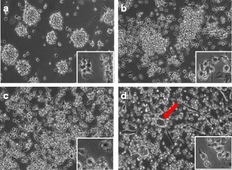
Morphology of eqMoDC at day 2 at 10× (large pictures) and 40× magnification (small inlaid pictures). Cells were incubated in the presence of HS (a), FBS batch S0113 (b), FBS batch S0613 (c) and FBS batch A15-101 (d), respectively. Pictures are representative of cells from 6 tested horses. The arrow in (d) indicates giant cells, presumably after uptake of apoptotic material, only seen in the FBS A15-101 condition