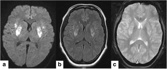
Fig. 1.

Brain magnetic resonance image (MRI) showed abnormal signal intensity in the bilateral putamen and anterior insular cortices. a diffuse restriction in the bilateral putamen and anterior insular cortices in diffusion weighted image, b high signal intensities in T2-weighted image on the same region, c: focal hemorrhage was not detected in susceptibility weighted image
