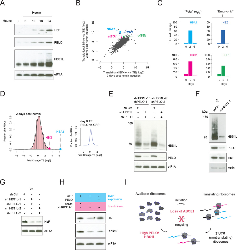
Fig. 5. PELO/HBS1L regulate ribosome homeostasis.
A. Hemoglobin, PELO, and HBS1L expression during the first 24 hours of hemin induction. B. Global translational efficiency (TE) changes after 2 days of hemin induction. C. TE changes for mRNAs encoding hemoglobin subunits measured two days post-induction. D. Histogram of TE changes for all mRNAs after 2 days of hemin treatment. Inset: TE ratio (PELO vs GFP) prior to Hemin induction. E. shRNA knockdown of PELO and HBS1L with two different shRNA hairpin sequences each. F and G. Hemoglobin expression after hemin induction is reduced by knock-down of HBS1L, enhanced by knock-down of PELO. H. RPS19 knockdown reduces hemin-induced hemoglobin accumulation, suppressed by overexpression of PELO. I. Model showing the loss of ABCE1 in differentiating K562 cells accompanied by induction of PELO/HBS1L to maintain ribosome homeostasis.

