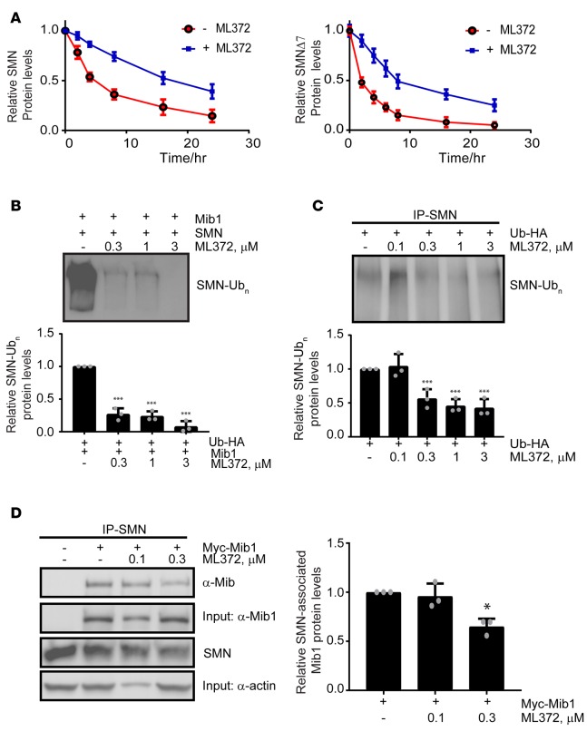
Figure 2. SMN ubiquitination is modulated by ML372.
(A) Pulse-chase analysis of myc-SMN (left panel) and myc-SMNΔ7 (right panel) transiently expressed in HEK-293T cells in the presence and absence of 0.3 μM ML372. (B) Recombinant SMN was incubated with the ubiquitin-activating enzyme (E1), the ubiquitin-conjugating enzyme (UBCH5b), and Mib1 with or without ML372 at the indicated concentrations. Densitometry analysis is shown as the mean ± SEM (n = 3, ***P < 0.001) (bottom panel). (C) HEK-293T cells were transiently transfected with 1 μg of HA-ubiquitin. Cells were treated with various concentrations of ML372 for 48 hours and ubiquitinated SMN was immunoprecipitated. Densitometry analysis is shown as the mean ± SEM (n = 3, ***P < 0.001) (bottom panel). (D) HEK-293T cells were transiently transfected with 1 μg of myc-Mib1. Cells were treated with various concentrations of ML372 for 48 hours, and SMN was immunoprecipitated. Western blot was used to determine SMN-associated Mib1. Densitometry analysis is shown as the mean ± SEM (n = 3, *P < 0.05) (right panel). P values were determined by 1-way ANOVA followed by Dunnett’s multiple comparisons test.

