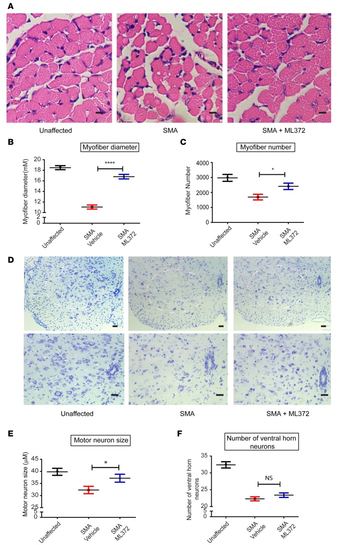
Figure 4. ML372 increases myofiber size and number and augments ventral horn neuron size.
SMNΔ7 spinal muscular atrophy (SMA) mice were treated with vehicle or ML372 from postnatal day 5 (PND5) to PND9. Black, unaffected; Red, SMNΔ7 SMA mice treated with vehicle; Blue, SMNΔ7 SMA mice treated with ML372. (A) H&E staining of tibialis anterior (TA) muscles from unaffected, SMA, and drug-treated SMA mice. Scale bars: 100 μm. (B) Mean myofiber diameter was compared in SMA and ML372-treated SMA mice. The results are indicated as the mean ± SEM (n = 20, ****P < 0.0001). (C) Myofiber numbers were counted in the 3 groups. The analysis is shown as the mean ± SEM (n = 20, *P < 0.05). (D) Nissl-stained cross-sections of spinal cord showing ventral horn neurons. Scale bars: 100 μm (upper and lower panels magnification ×10 and ×20, respectively). (E) The size of motor neurons was analyzed in the 3 groups and the mean ventral horn neuron size was compared in SMA mice treated with vehicle or ML372. The data are shown as the mean ± SEM (n = 20, *P < 0.05). (F) The number of motor neurons was determined in the 3 groups, and the analysis is shown as the mean ± SEM (n = 20). P values were determined by unpaired 2-tailed Student’s t test.

