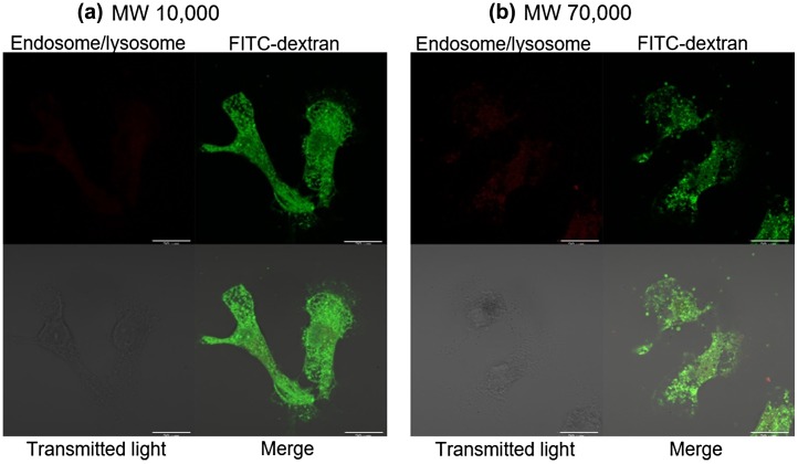
Figure 4.
Confocal microscopy of cells 24 h after fET in the presence of (a) FITC-dextran10,000 or (b) FITC-dextran70,000.
Cells were treated with fET (0.34 mA cm–2, 15 min) in the presence of FITC-dextran solution. After 23.5 h, the cells were stained with LysoTracker Red DND for 0.5 h, then observed with a confocal laser scanning microscope. The scale bars indicate 20 μm.

