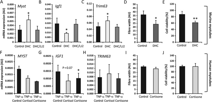
Figure 4.

Levels of mRNA in primary cultures of differentiated myotubes isolated from quadriceps muscle biopsies from WT mice (A–C) and human quadriceps biopsies (F–H) (n = 3 per variable), determined by RT‐qPCR. To induce 11β‐HSD1 expression, human primary cultures were pretreated with TNF‐α (10 ng/ml) for 48 h prior to a 12‐h washout. Cells were then incubated for 16 h with either control, active cortisol (100 nmol/l), or its inactive precursor cortisone (100 nmol/l). In murine culture, cells were treated for 24 h with either control medium, the inactive corticosterone precursor DHC (100 nmol/l), or DHC in combination with the selective 11β‐HSD1 inhibitor LJ2 (1000 nmol/l). Fibre width and cell viability were determined with Image J analysis software and MTT assay, respectively, in murine (D, E) and human (I, J) primary myotubes following incubation with normal control medium and DHC (100 nmol/l)‐containing medium over a period of 5 days (n = 3 per variable). Values are expressed as mean ± SE. Statistical significance was determined with one‐way anova with a Dunnett post hoc analysis. *p < 0.05, **p < 0.005.
