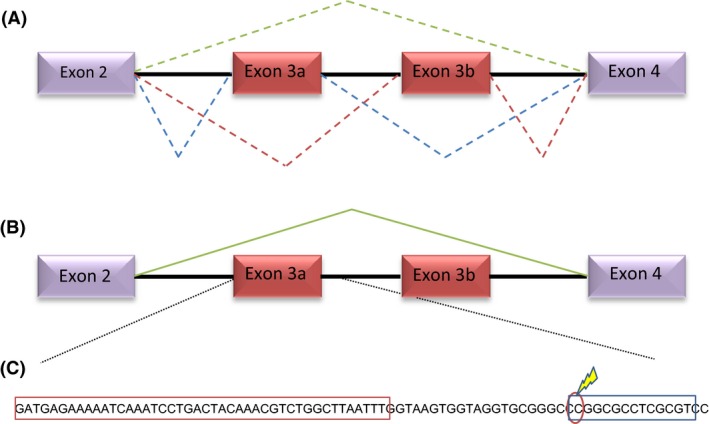
Figure 2.

Patterns of splicing and DNA methylation of the Tuta absoluta nAChR α6 subunit gene. (A) The spinosad susceptible strain Spin exhibits mutually exclusive splicing of exon 3a/3b (blue and red dashed lines), but a low frequency of transcripts exhibits exon skipping (green dashed line). (B) In the spinosad resistant strain SpinSel, all Tα6 transcripts exclude both exons 3a and 3b (green solid line). (C) Nucleotide sequence of exon 3a and immediate region downstream highlighting the position of the single CpG site that is 30% methylated in the SpinSel strain (marked with a lightening bolt). The sequence encoding exon 3a is boxed in red. The region boxed in blue indicates a predicted CTCF binding site. [Colour figure can be viewed at wileyonlinelibrary.com]
