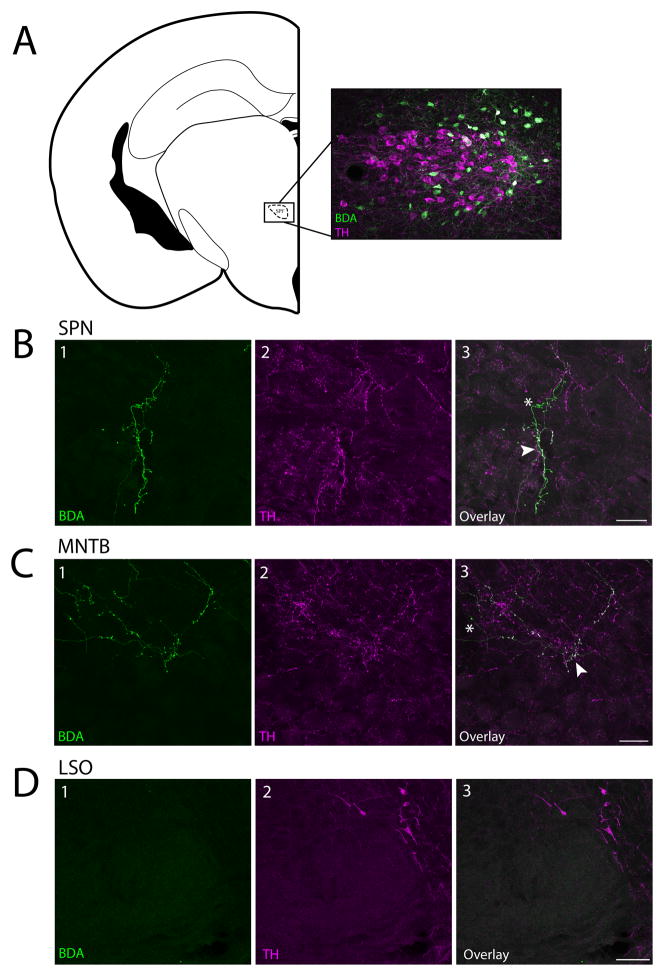
Figure 1. Dopaminergic cells in the SPF project to the SPN and MNTB but not the LSO.
A: Coronal section showing a BDA deposit (green cell bodies) in the SPF. TH-positive cells are shown in magenta. B: Fibers in the SPN were anterogradely labeled and TH-positive. B1: BDA labeled fibers. B2: TH-positive fibers. B3: Overlay. C: Fibers in the MNTB were anterogradely labeled and TH-positive. C1: BDA labeled fibers. C2: TH-positive fibers. C3: Overlay. D. There were no anterogradely labeled fibers in the LSO. B–D: Arrows point to examples of anterogradely labeled fibers colocalized with TH, asterisks mark TH-negative anterogradely labeled fibers. Coronal section modified from Paxinos and Franklin (2001). Scale bar: A, D 100μm; B, C 25μm. Abbreviations: SPF, subparafascicular thalamic nucleus; SPN, superior paraolivary nucleus; MNTB, medial nucleus of the trapezoid body: LSO, lateral superior olive.

