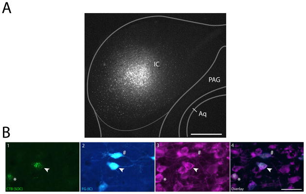
Figure 5. Single neurons in the SPF project to both the IC and SOC.
A: Fluorogold deposit in the IC, with an overlaid schematic coronal section based off of Paxinos and Franklin (2001). B1: Retrogradely labeled cells in the SPF after CTB deposit in the MNTB (deposit site shown in Fig. 3A). B2: Retrogradely labeled cells in the SPF after IC deposit. B3: TH-positive cells in the SPF. B4: Merged view showing colocalization of tracer and TH. Arrows show a TH-positive cell retrogradely labeled from IC and MNTB deposits. Asterisk marks a TH-positive cell that is retrogradely labeled from only the MNTB. Pound sign marks a TH-positive neuron that is retrogradely labeled only from the IC. Scale bar: A 250μm; B 40μm. Abbreviations: CNIC, central nucleus of inferior colliculus; ECIC, external cortex of inferior colliculus; DCIC, dorsal cortex of inferior colliculus; PAG, periaqueductal gray; Aq, aqueduct.

