Fig. 6.
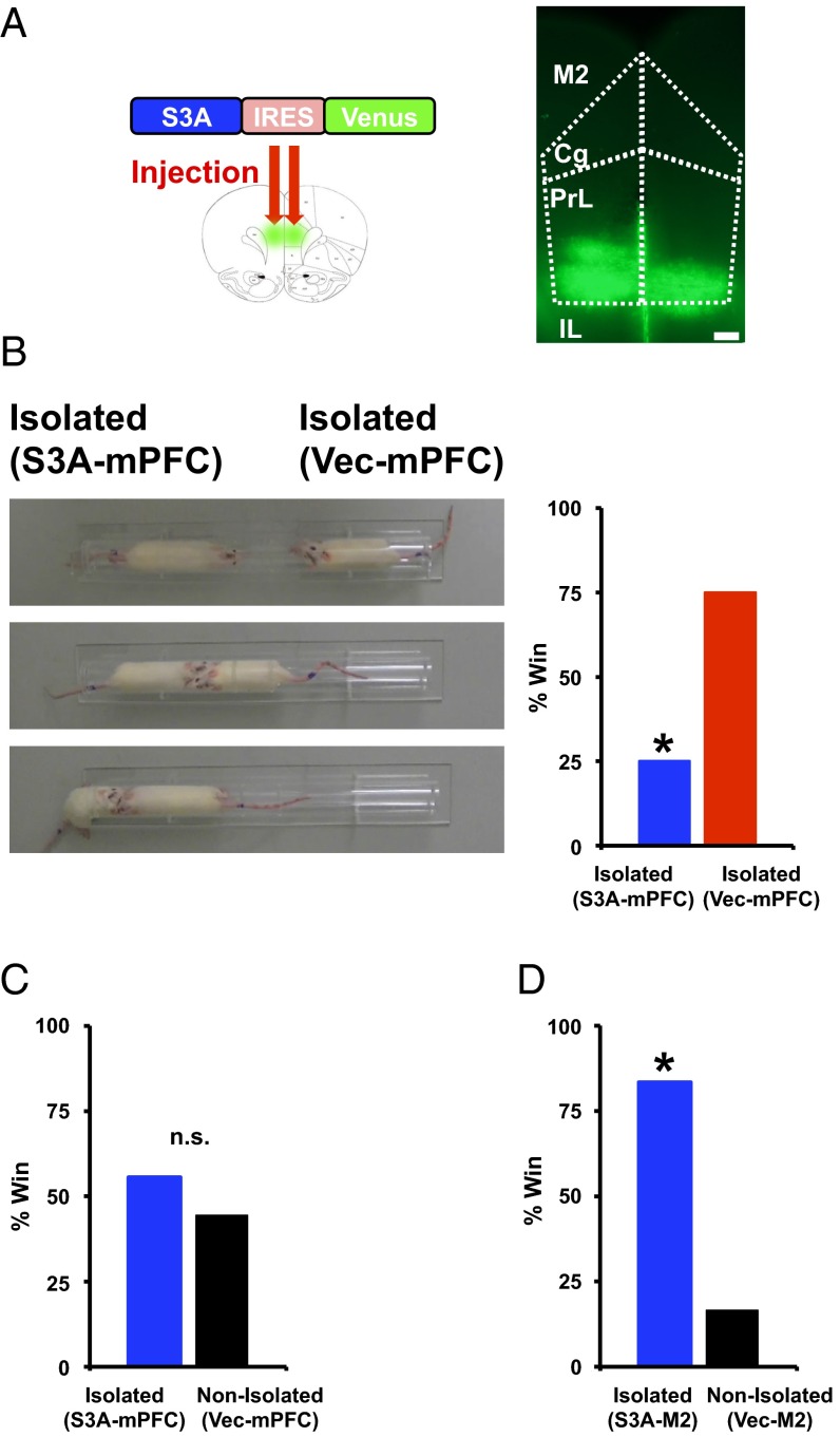
Activation of ADF/cofilin in the mPFC suppresses the enhancement of social dominance in socially isolated rats. (A) Example of brain slices taken from an animal injected with Lenti-S3A-IRES-Venus. Subregion boundaries are indicated with dotted lines. (Cg, cingulate cortex; IL, infralimbic cortex; M2, secondary motor cortex; PrL, prelimbic cortex). (Scale bar, 300 μm.) (B) Social dominance tube test of socially isolated rats with mPFC injection of ADF/cofilin S3A (S3A-mPFC). (Left) Captured video images from a representative match. From top to bottom, the beginning to the end of the match is sequentially indicated. The socially isolated rat expressing S3A (mPFC) was pushed out by the socially isolated rat expressing vector in the mPFC (Vec-mPFC). (Right) Percentage of wins in the matches between socially isolated rats expressing S3A (mPFC) and vector (mPFC) (eight matches). (C) Social dominance tube test between socially isolated rats expressing S3A (mPFC) and nonisolated rats expressing vector (mPFC). Isolated rats (S3A-mPFC) exhibited social dominance comparable to that of nonisolated animals (vector-mPFC) (nine matches), (D) but not rats with other brain area, M2 motor cortex, injection (six matches). *P < 0.05 (χ2 test, B, C, and D). n.s., not statistically significant.

