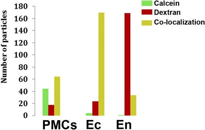
Fig. 4.
The size, type of labeling, and location of 522 particles, obtained from analysis of individual Lightsheet images (Materials and Methods), similar to the slices shown in Fig. 3 D–F. The green columns represent particles that are only labeled with calcein. The red columns represent particles that are only labeled with alexa-dextran. The yellow columns represent particles in which calcein and alexa-dextran labels colocalize. Approximately half of the particles in the PMCs display label colocalization. Almost all of the particles that are located inside ectoderm cells (Ec) display label colocalization. Most of the particles that are located inside endoderm cells (En) only display alexa-dextran labeling.

