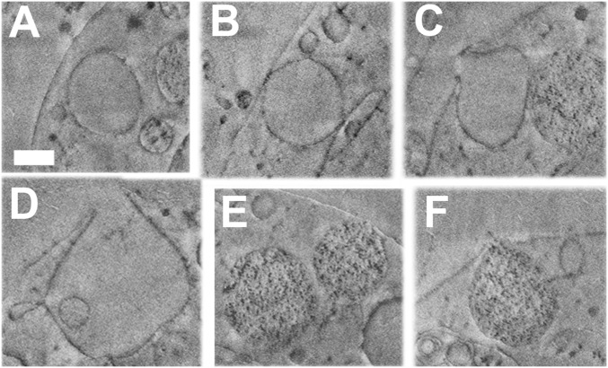
Fig. 5.
Cryo-FIB-SEM micrographs of parts of a PMC from a sea urchin larva at the prism stage. Proteins, lipids, and membranes appear dark, whereas water-rich regions such as aqueous cytosol appear in uniform light gray in cryo-FIB-SEM. The series of micrographs is taken from Movie S1. (A–D) Vesicles bearing a uniform content similar in texture and gray levels to cytoplasm and extracellular fluids. (E and F) Vesicles containing dark particles. (A) No contact of the vesicle with the plasma membrane. (B) The vesicle contacts the plasma membrane. (C) The vesicle is open toward the blastocoel. (D) The vesicle has a wide opening toward the blastocoel and branches into smaller vesicles. (E) One vesicle (to the right) touches the plasma membrane, but the other does not. (F) Vesicle in contact with the plasma membrane and open toward the blastocoel. (Scale bars: 500 nm for all panels.)

