Fig. 1.
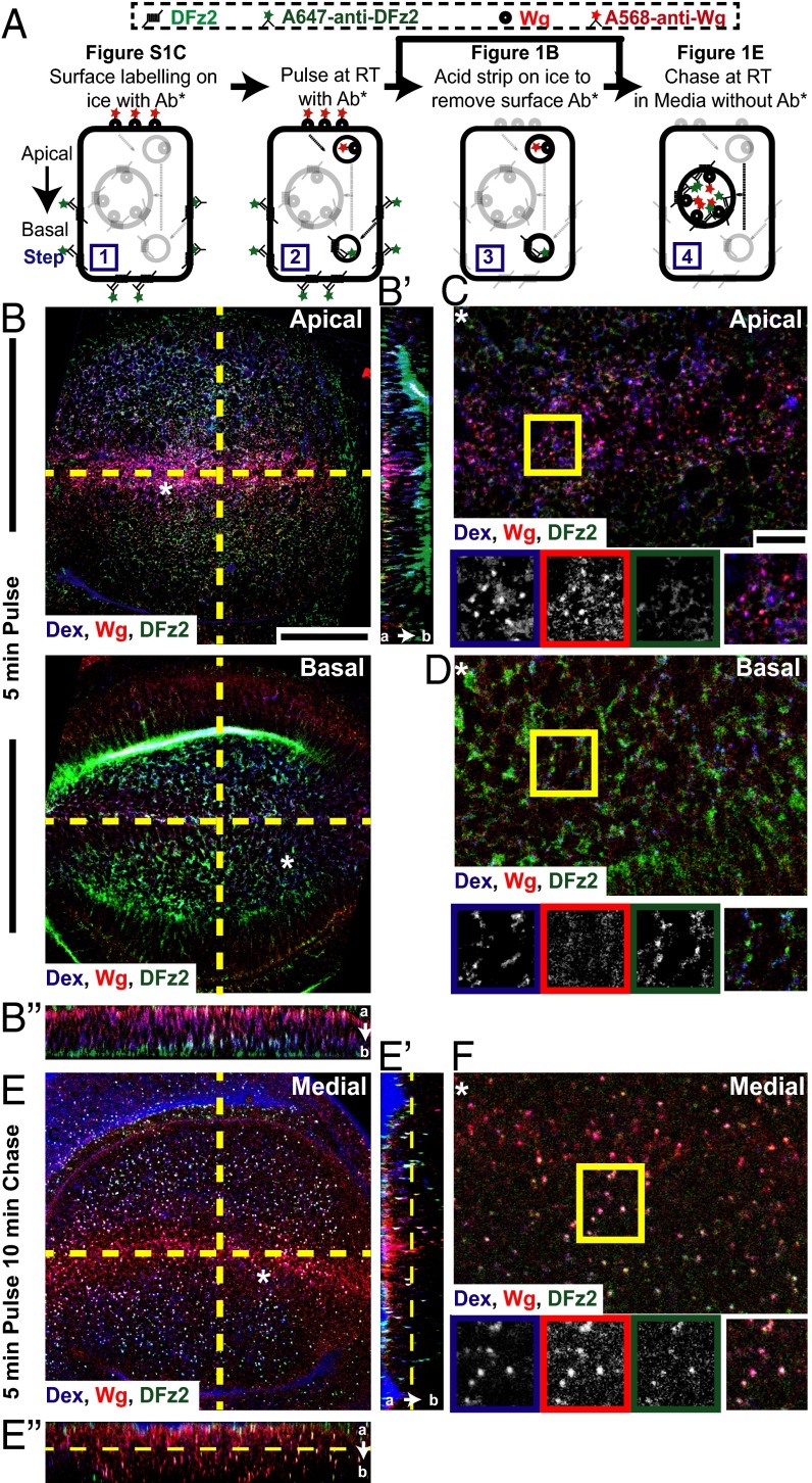
Wg is endocytosed apically devoid of its signaling receptor, DFz2. (A) Endocytic assay: Surface distribution of Wg and DFz2 evaluated by incubating fluorescently labeled primary antibodies (Ab*) A568-anti-Wg, A647-anti-DFz2 on ice (step 1) (Fig. S1C). Surface-labeled wing discs are incubated at room temperature for 3–5 min with Ab* and Dex to image early endosomes (step 2) and acid washed to remove the Ab* left at the surface (step 3; see B–D), followed by chase in media without Ab* (step 4; see E and F). (B–F) Apical (B, Upper) and basal (B, Lower) confocal sections of control (w1118) wing discs shows “5-min pulse” endosomes (B) and the medial plane of w1118 wing discs shows “5-min pulse and 10-min chase” endosomes (E) of Dex (blue), Wg (red), and DFz2 (green). YZ (B′/E′) and XZ (B″/E″) sections (along yellow lines in B and E) are oriented apical to basal (a → b). Regions (*) from B and E are magnified in C, D, and F. Boxed regions (C, D, and F) are magnified (∼1.5×) and shown below with indicated probes in the color outlines. Images have been rotated (B–D), background-subtracted, and median-filtered with intensities appropriately scaled. Wg and Dex colocalize extensively in apical endosomes, whereas DFz2 endosomes are predominantly found basally, yet separate from Dex endosomes 5-min postinternalization. With time, all three colocalize in a common endosome in more medial planes. (Scale bars, 50 μm in B and E and 10 μm in C, D, and F.)

