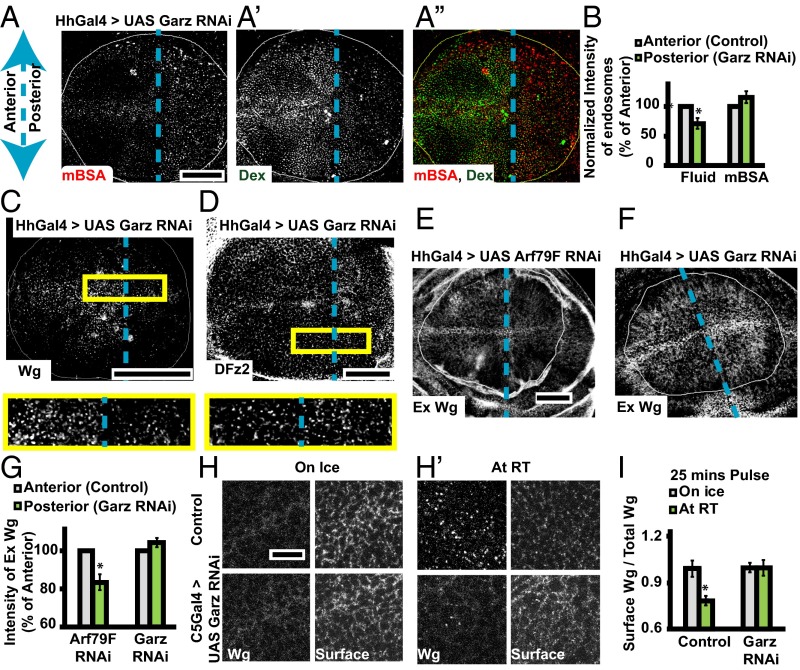
Fig. 3.
Garz perturbation specifically affects fluid phase and Wg endocytosis. Dashed blue line indicates the anterior/posterior (A/P) compartment boundary. Posterior is to the right in all disc images. (A) Confocal projections of wing discs with HhGAL4 driving Garz RNAi in the posterior showing: 15-min endocytosis of CD cargo–mBSA (A; red in merge), Dex (A′; green in merge), and merge (A″); 10-min endocytosis of Wg (C; Dex uptake in Fig. S3I) and DFz2 (D; Dex uptake in Fig. S3J). Histogram in B represents the normalized (to the compartment area) intensity of Dex and mBSA endosomes in the posterior with respect to the anterior (n = 6 wing discs). Yellow boxes (C and D) are magnified (∼2.5×) below. Images are average intensity projections of confocal planes (13 planes in A; 3 apical planes in C; and 3 basolateral planes in D; depth, 1.0 μm). Although Wg and fluid (Dex) endocytosis is severely reduced in the posterior with respect to the anterior, DFz2 and mBSA endocytosis is not. (E–G) Confocal projections (six to eight confocal planes; depth, 1.0 μm) of extracellular Wg staining in wing discs with Arf79F RNAi (E) or Garz RNAi (F) driven in the posterior compartment with HhGAL4. Histogram (G) shows that the surface levels of Wg is not reduced across A/P axis upon depletion of Garz, unlike Arf79F depletion. (H–I) Surface internalization assay (SI Materials and Methods) using A568 anti-Wg (1° antibody-Wg) and A647 anti-mouse (2° antibody-surface) on control wing discs (H, Upper) and those with Garz RNAi expressed under C5GAL4 (H, Lower): Confocal slices from wing discs maintained on ice/room temperature are represented as H/H′. Histogram in I shows the amount of 2° antibody bound after 25 min at the indicated temperature, normalized to the total 1° bound Wg initially present at the cell surface (SI Materials and Methods). n = 5 discs; 2 repeats; *P < 10−3. All images are background-subtracted, median-filtered (A, C, D) and intensities appropriately scaled. (Scale bars, 50 μm in A–A″ and C–F, and 10 μm in H and H′.)

