Fig. 4.
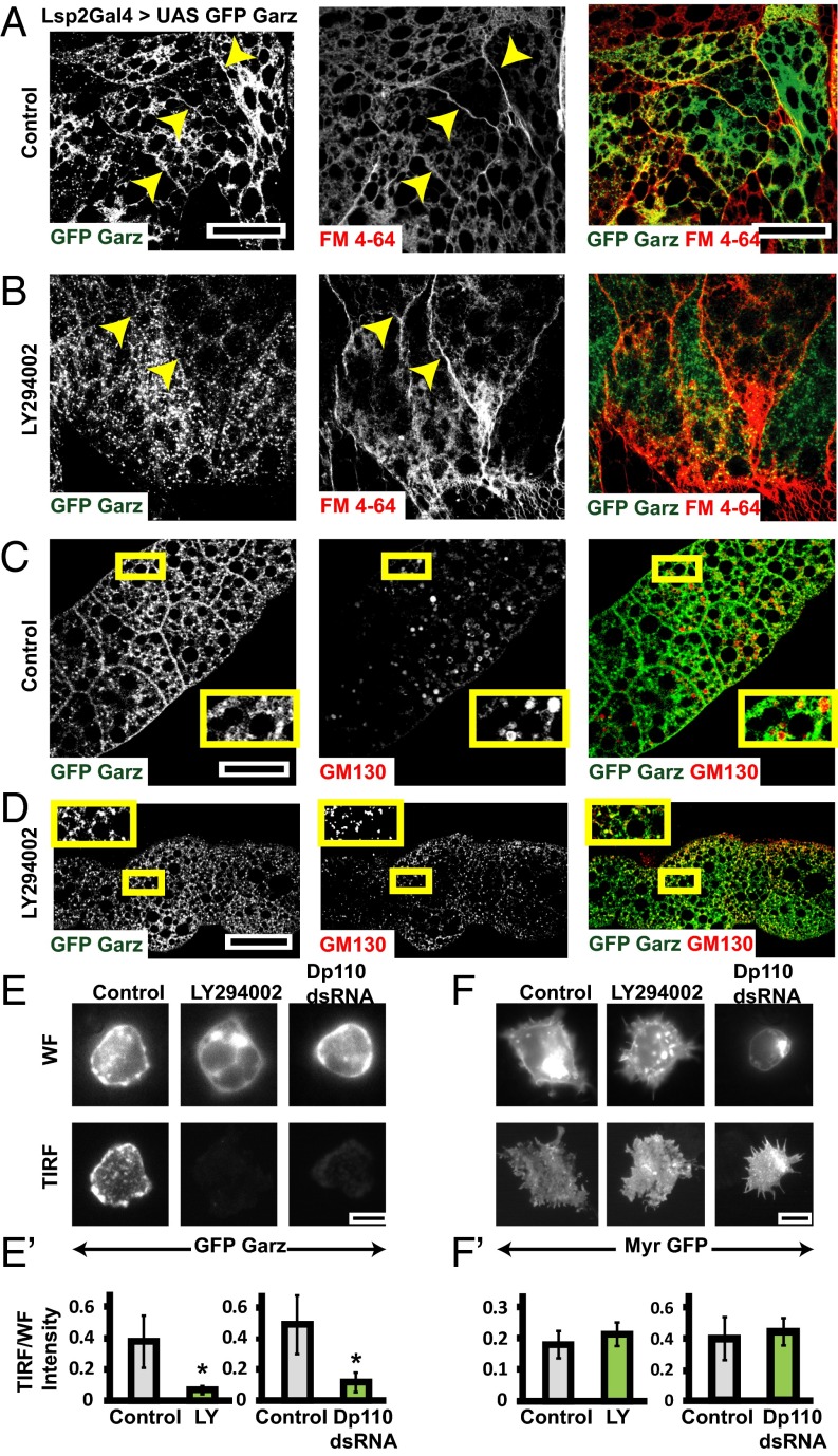
Class I PI3K mediates plasma membrane localization of Garz. (A–D) Confocal slices of UAS-GFP Garz (A–D; green) expressed in fat bodies using LSP2 GAL4 showing marked accumulation (A) along the plasma membrane labeled by FM4-64 dye (red) and (C) in Golgi labeled by GM130 (red). Upon treatment with LY294002 the plasma membrane localization of GFP–Garz is lost (B), but the Golgi localization appears unaffected (D). Yellow arrows in A and B point toward plasma membrane. The outlined regions in yellow are magnified (∼2.1×) and shown as Insets in C and D. (E and F) TIRF and widefield (WF) images of S2R+ cells expressing Actin GAL4 and pUAST-GFP–Garz (E) or pUAST-myristoylated-GFP (F) in untreated (Control) or cells treated with LY294002 or Dp110 dsRNA. The ratio of TIRF/WF, which reflects the relative extent of membrane localization of the protein, is plotted in E′ and F′. Although the ratio of GFP–Garz reduces in both the PI3K perturbed conditions (*P < 0.2 × 10⁻³), the ratio of myr GFP (although these cells are more spread with many filopodia) is not significantly different between the wild-type and perturbations. All images are background-subtracted and intensities appropriately scaled for representation. (Scale bars, 50 μm in A–D and 10 μm in E and F.)

