Fig. 5.
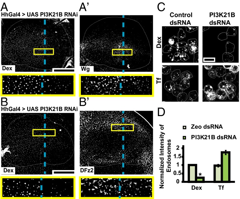
PI3K perturbation specifically affects fluid phase and Wg endocytosis. (A and B) Confocal images of wing discs expressing PI3K21B RNAi using HhGAL4 in the posterior compartment showing 10-min endosomes of (A and A′) Dex and Wg, and (B and B′) Dex and DFz2, probed using FITC-Dex and A568 anti-Wg or A647 anti-DFz2. Both Wg and Dex uptake is reduced in the posterior compartment compared with the control anterior side, whereas DFz2 endosomes are similar in the posterior compared with anterior. Images are average intensity projections of confocal planes (10 apical planes in A; 7 apical planes in B; 7 basolateral planes in B′), each of depth 1.0 μm with background-subtraction and median-filtering and intensities appropriately scaled for representation. The outlined regions in yellow are magnified (∼3.7×) and shown below respective images. Posterior compartment is to the right in all wing discs. Dashed blue line approximately indicates the A/P compartment boundary. (C and D) WF images of S2R+ cells with control (Zeocin) or PI3K21B dsRNA showing 10-min uptake of Dex (C, Upper) and transferrin (C, Lower). Dex uptake is reduced whereas transferrin uptake is slightly enhanced in PI3K21B dsRNA cells as quantified in the histogram (D). *P < 10−20 obtained from 80 to 100 cells with two replicates. Images are background subtracted and intensities are appropriately scaled for representation. (Scale bar, 50 μm in A, A′, B, and B′ and 10 μm in C.)

