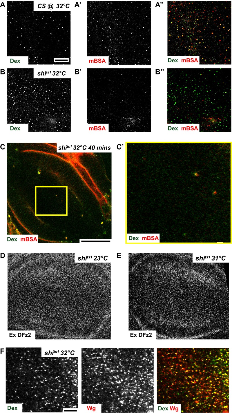
Fig. S2.
Shibire is not required for Wg endocytosis (supplementary to Fig. 2). (A and B) High-resolution confocal images of 10-min pulsed endosomes of CG marker, Dex (green in merge) and CD cargo mBSA (red in merge), in Control (CS) and shits1 wing discs upon shifting to restrictive temperature (32 °C) for a total of 15 min. Although mBSA endocytosis is abrogated in shits1 wing discs (B′), Dex uptake (B) proceeds normally. (C) Z projection of confocal slices of wing discs depicting Dex and mBSA endocytosis in shits1 wing discs maintained at restrictive temperature (32 °C) for a total of 40 min. Yellow box in C is magnified (∼3.4×) in C′. Note that both Dex (CG) and DFz2 (CD) endocytosis is severely affected upon prolonged incubations at restrictive temperatures. (D and E) Confocal images with surface staining of DFz2 in control (shits1 maintained at room temperature, as shown in Fig. 2A) and shits1 wing discs after shifting to restrictive temperature for 15 min (E; as shown in Fig. 2B), indicating that although extracellular DFz2 is present, DFz2 endocytosis is inhibited upon perturbation of Shibire. (F) High-resolution confocal images of 5- to 8-min pulsed endosomes of Dex (green in merge) and Wg (red in merge) in shits1 wing discs after shifting to restrictive temperature (32 °C) for a total of 15 min, demonstrating that both Dex and Wg uptake is unaffected upon perturbation of Shibire and continued to appear in colocalized endosomes. All images are background-subtracted and intensities are appropriately scaled for representation. (Scale bars, 10 μm in A, A′, A″, B, B′, and B″ and F and 50 μm in C. Scaling in D and E is the same as in Fig. 2 A and B, respectively.)

