Fig. S4.
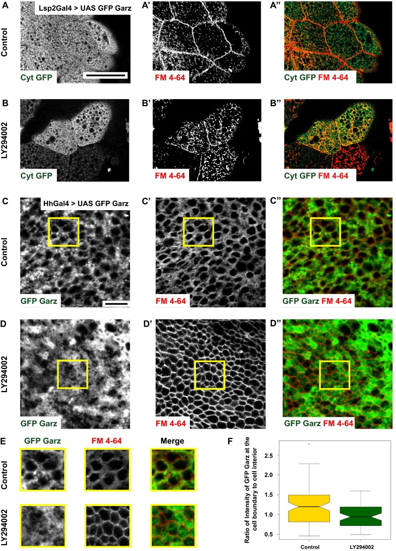
Class I PI3K mediates plasma membrane localization of Garz (supplementary to Fig. 4). (A and B) Confocal sections of cytosolic GFP (A; green in A″) overexpressed in fat bodies of Drosophila using LSP2-GAL4 shows the distribution of GFP with no enrichment at the plasma membrane marked by FM4-64 dye (A′; red in A″). The distribution is unchanged upon treatment with LY294002 inhibitor (B). (C–E) GFP–Garz (C; green in C″) in wing disc expressed using Hh-GAL4 shows enrichment along the plasma membrane boundary labeled by FM4-64 dye (C′; red in C″). Note that upon treatment with LY294002 (D), GFP–Garz appears more diffused (D, D″). Outlined ROIs (yellow) are magnified (∼1.5×) and shown in E. Histogram (F) shows the ratio of GFP–Garz intensity at the cell boundary and cell interior quantified in control and LY294002 treated wing discs (SI Materials and Methods). This ratio is reduced upon inhibitor treatment indicative of reduced localization of GFP–Garz to the plasma membrane. All images are background-subtracted and intensities are appropriately scaled for representation. (Scale bars, 50 μm in A and B and 10 μm in C and D.)

