Fig. S7.
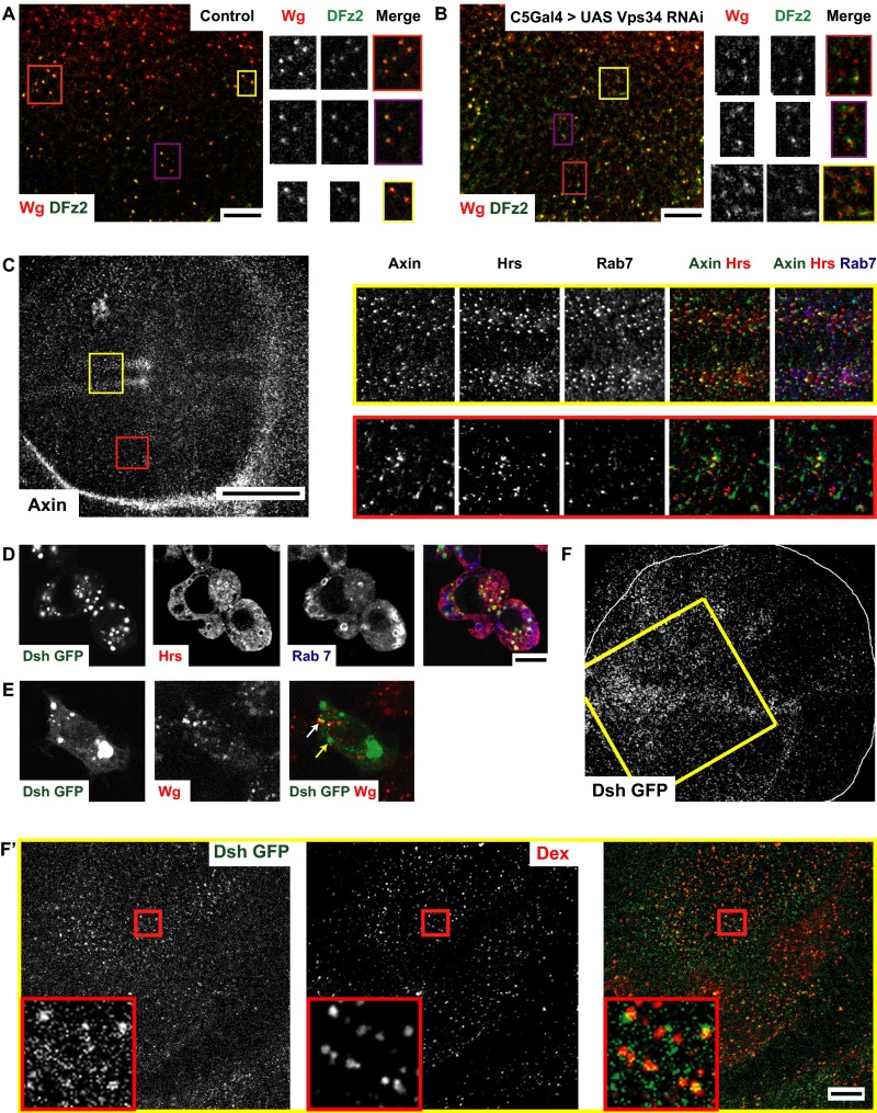
Wg signal transduction components are localized to endosomes (supplementary to Fig. 7). (A and B) Confocal sections of control (A; C5GAL4/+) or C5GAL4 driving Vps34 RNAi (B) wing discs pulsed for 5 min and chased for 10 min to visualize Wg and DFz2 endosomes. Note depletion of Vps34 causes defects in the merging of Wg (red) and DFz2 (green) endosomes compared with wild-type wing disc; Vps34 RNAi depleted discs show many segregated early endosomes of Wg and DFz2 at the end of the chase period. The yellow outlined regions in A and B are magnified (∼2×) and represented as a montage showing the grayscale intensities in each channel along with the merge. (C) Confocal sections of w1118 wing discs immunostained for Axin (gray in full disc; green in merge), along with endosomal markers Hrs (red in merge) and Rab7 (blue in merge). Axin shows a predominantly punctate distribution. Outlined regions in yellow (apical planes) and red (basal planes) in C are magnified (∼3×) and represented as a montage showing the gray-scale intensities in each channel along with the merge. Note: Axin colocalizes extensively with Hrs and Rab7. 29.27 ± 3.02% of anti-Axin staining colocalized with Hrs and 20.91 ± 1.82% of anti-Axin colocalizes with Rab7 (n = 4 wing discs). (D and E) S2R+ cells transfected with Dsh GFP (expressed under its own promoter; green in merge) is (D) immunostained with Hrs (red in merge) and Rab7 (blue in merge) or (E) cultured with secreted Wg and subsequently labeling the 5-min Wg endosomes using A568 anti-Wg (red in merge). Dsh accumulates in long-lived vesicular structures enriched in Hrs but not that prominantely in Rab7 [57.93 ± 10.39% of Dsh GFP colocalizes with Hrs, whereas only 11.55 ± 8.24% colocalizes with Rab7 (n = 10 cells)] and Dsh-GFP also labels early endosomes containing Wg (white arrow) in addition to the large vesicles (yellow arrow) as seen in D; 44.96 ± 8.54% of Wg endosomes (5 min) strongly colocalizes with Dsh-GFP(n = 8 cells). (F and F′) Dsh-GFP (green in merge) is loaded onto early endosomes outlined by 10-min uptake of Dex (red in merge) in wing discs expressing Dsh-GFP under its native promoter in addition to its diffused distribution in the subapical planes. 50.016 ± 3.06% of early endosomes containing TMR-Dex colocalizes with Dsh-GFP punctae (n = 4 wing discs). Regions outlined in yellow, adjacent to the dorsoventral boundary, are further magnified (∼1.6×) in F′. All images are background subtracted with intensities appropriately scaled for representation. (Scale bars, 50 μm in C and F and 10 μm in A, B, D, E, and F′.)

